NSJ Bioreagents

Many products are offered at multiple sizes to better fit researchers testing needs and budget constraints. Comprehensive product data sheets provide product information and testing data images and are backed up by experienced technical service. And all products are 100% guaranteed to work as stated on the product data sheets.

Here is the best sales products which are used by many researchers all over the world.
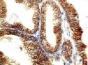
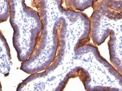
PD-L1 Antibody / B7-H1 / CD274 [clone PDL1/2746] (V3955)
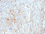
Engagement of CD28 by B7-1 (CD80) or B7-2 (CD86) in the presence of antigen promotes T-cell proliferation, cytokine production, differentiation of effector T-cells and the induction of BCLX, a promoter of T-cell survival. recruitment of CTLA4 by B7-1 or B7-2, on the other hand, may inhibit proliferation and interleukin-2 (IL-2) production.
PD-L1 is 290-amino acid type I transmembrane protein, which is 20% and 15% identical to B7-1 and B7-2, respectively, has immunoglobulin V-like and C-like domains and a 30-amino acid cytoplasmic tail. PD-L1 does not bind CD28, cytotoxic T-lymphocyte A4 or ICOS (inducible co-stimulator). IL-2, although produced in small amounts, is required for the effect of PD-L1 co-stimulation. PD-L2 protein contains a signal sequence, IgV- and IgC-like domains, a transmembrane region and a cytoplasmic region.
Constitutive expression of PD-L1 and PD-L2 on parenchymal cells of heart, lung and kidney suggests that the PD-1-PD-L system could provide unique negative signaling to help prevent autoimmune diseases.
Please check Product data sheet for the detail of this product.
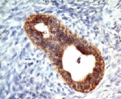
Histone Antibody / Histone H1 [clone 1415-1] (V2567)
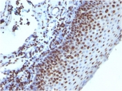
Eukaryotic histones are basic and water-soluble nuclear proteins that form hetero-octameric nucleosome particles by wrapping 146 base pairs of DNA in a left-handed super-helical turn sequentially to form chromosomal fiber. Two molecules of each of the four core histones (H2A, H2B, H3, and H4) form the octamer; formed of two H2A-H2B dimers and two H3-H4 dimers, forming two nearly symmetrical halves by tertiary structure. Over 80% of nucleosomes contain the linker Histone H1, derived from an intronless gene that interacts with linker DNA between nucleosomes and mediates compaction into higher order chromatin.
Histones are subject to posttranslational modification by enzymes primarily on their N-terminal tails, but also in their globular domains. Such modifications include methylation, citrullination, acetylation, phosphorylation, sumoylation, ubiquitination and ADP-ribosylation.
Please check Product data sheet for the detail of this product.
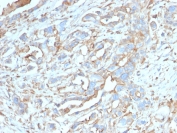
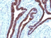
E-Cadherin Antibody [clone CDH1/1525] (V3190)
Recognizes a protein of 80-120kDa, identified as E-cadherin. Cadherins comprise a family of Ca2+-dependent adhesion molecules that function to mediate cell-cell binding critical to the maintenance of tissue structure and morphogenesis. The classical cadherins, E-, N- and P-cadherin, consist of large extracellular domains characterized by a series of five homologous NH2 terminal repeats. The relatively short intracellular domains interact with a variety of cytoplasmic proteins, such as β-catenin, to regulate cadherin function.
E-cadherin plays an important role in epithelial cell adhesion. A decreased expression of E-cadherin is associated with metastatic potential and poor prognosis in breast cancer, prostate and esophageal cancer. In combination with p120 Catenin, it is useful for the differentiation between ductal (E-cadherin +) and lobular (E-cadherin -) breast carcinomas. It may also help in diagnosis of mesothelioma.
Please check Product data sheet for the detail of this product.
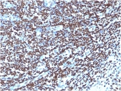
EpCAM Antibody / Cytoplasmic domain [clone EGP40/1110] (V2686)
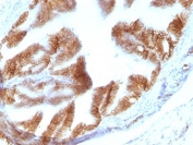
EGP40 is a 40-43kDa transmembrane epithelial glycoprotein, also identified as epithelial specific antigen (ESA), or epithelial cellular adhesion molecule (Ep-CAM). It is expressed on baso-lateral cell surface in most simple epithelia and a vast majority of carcinomas.
This antibody has been used to distinguish adenocarcinoma from pleural mesothelioma and hepatocellular carcinoma. This antibody is also useful in distinguishing serous carcinomas of the ovary from mesothelioma.
Please check Product data sheet for the detail of this product.
EMA Antibody / MUC1 / Mucin-1 [clone MUC1/845] (V2371)
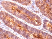
This antibody recognizes proteins in MW range of 265-400 kDa, identified as different glycoforms of MUC1 (Mucin-1) or EMA (epithelial membrane antigen). The alpha subunit has cell adhesive properties. It can act both as an adhesion and an anti-adhesion protein.
MUC1 / Mucin-1 may provide a protective layer on epithelial cells against bacterial and enzyme attack. The beta subunit contains a C-terminal domain, which is involved in cell signaling through phosphorylations and protein-protein interactions. In immunohistochemical assays, the MUC1 / EMA antibody superbly stains routine formalin/paraffin carcinoma tissues. MUC1 antibody is useful as a pan-epithelial marker for detecting early metastatic loci of carcinoma in bone marrow or liver.
Please check Product data sheet for the detail of this product.
List of antibody categories
- ABO
- Angiogenesis
- Apoptosis
- Apoptosis Marker
- Astrocyte
- Autophagy
- B-Cell
- Beta Cell
- Calcium Channel
- Cancer
- Cardiovascular
- Cell Biology
- Cell Cycle
- Cellular Marker & Epitope Tag
- Cytokine
- Dendritic Cell
- Developmental Biology
- Endoplasmic Reticulum
- Endosome
- Endosome Marker
- Endothelial
- Endothelial Cell
- Endothelial Cell Marker
- Epigenetics
- ER
- Erythrocyte
- Follicular Lymphoma
- Granuloctye
- Hairy Cell Leukemia
- Hepatocellular Carcinoma
- Hepatocyte
- Hepatocyte Marker
- Human Protein Microarray Validated
- Ig
- Immunology
- Innate Immunity
- Isotype Control
- Late Endosome Marker
- Leukocyte
- Loading Control
- Lymphocyte
- Macrophage
- Mantle Cell
- Melanoma
- Mesenchymal Cell
- Metabolism
- Microbiology
- Microglia
- Mitochondria
- Mitochondrial
- Monocyte
- Myeloid Cell
- Neuronal
- Neuronal Marker
- Neuroscience
- Neutrophil
- NF-kB
- NK Cell
- Nuclear
- Phospho
- Platelet
- Popular
- Proliferation
- Rabbit Monoclonal
- Recombinant
- Recombinant Mouse
- Renal Cell
- Signal Transduction
- Skeletal Muscle
- Smooth Muscle
- Stem Cell
- Stem Cell Marker
- T-Cell
- Thyroid
- TLR & Related
- Zinc Transporter
Protocols
Please contact us for any inquiries, questions, or information requests.
Tokyo Future Style, Inc.
info@tokyofuturestyle.com
TEL:029-851-9222 FAX:029-851-9220
